


 A basic understanding of skin anatomy is important when explaining the process of skin biopsy. Each component of the skin plays a role in its daily function, therefore every component is a source of vital information that can be captured and assessed with a skin biopsy.
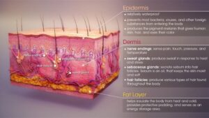
Below are a few of the basic components of skin followed by a brief description their functions.
- Hair – Hair serves a protective role in the skin. On most locations of the body, hair offers a protective covering, which regenerates on a regular basis. In some places, hair serves as a filter (such as in the nose and ears), a moisture and heat retention mechanism (such as the armpits and genital region), and in the middle ear it serves as a mechanism for regulating balance. Each hair follicle (in the hairy parts of the skin) is attached to a muscle, the arrector pili (see Arrector Pili for more information).
- Stratum Corneum – This is the dead skin layer that is visible when you look at your skin. It functions to protect the living cells beneath by providing a hard barrier between the outside world and the delicate cells inside. The stratum corneum is useful for diagnosis because in some conditions the stratum corneum will become thinner than normal.
- Epidermis – The epidermis is the next layer under the stratum corneum. Its function is to protect the body. It produces cells that will eventually become stratum corneum cells. It contains sensory nerves specifically small diameter sensitive temperature fibers. It is these sensory nerves that are helpful when evaluating a skin biopsy.
- Sensory Nerves – These are the nerves that innervate the epidermis. These nerves are the subject of evaluation when examining a skin biopsy after it has been immunostained. The sensory nerves in the epidermis serve to sense and transmit heat, pain, and other noxious sensations. When these nerves are not functioning properly they can produce sensations such as numbness, pins-and-needles, pain, tingling, or burning. When evaluated, characteristics of the nerve such as total number, contiguity, diameter, branching, swelling, and overall health are taken into consideration.
- Dermis – The dermis is the next layer under the epidermis. The dermis contains all of the other sub-epidermal structures mentioned below. Dermis is characterized by loose, ribbon-like cells that hold dermal structures in place and serves to contain fluids.
- Arrector Pili Muscle – This is a tiny muscle that attaches to the base of a hair follicle at one end and to dermal tissue on the other end. In order to generate heat when the body is cold, the arrector pili muscles contract all at once, causing the hair to “stand up straight” on the skin. The arrector pili muscle is a source of information when evaluating a skin biopsy since it is well-innervated with autonomic nerves that control when the muscle contracts. These autonomic nerves are also visible when the skin biopsy is immunostained.
- Sebaceous Glands – These structures are associated closely with hair follicles because they produce an oily substance that coats and protects the hair shaft from becoming brittle.
- Sweat Glands – These glands produce moisture (sweat) which is secreted through tiny ducts to the surface of the skin (stratum corneum). The moisture serves as a cooling agent by making the surface of the skin moist. This moisture then evaporates and lowers the temperature of the skin.
- Basket Cells – These structures surround the base of hair follicles and serve as pressure sensors. They are a source of valuable information when assessing overall nerve health and condition.
- Blood Vessels – These structures carry vital nutrients and oxygen-rich blood to the cells that make up the layers of skin and then carry away waste products. Often, the blood vessels are in close proximity to collections of nerve bundles in the dermal and subdermal layers.