


 Many different tests can be used to diagnose a patient with peritoneal carcinomatosis (or peritoneal mets) that started from another organ such as the appendix, colon, stomach, or ovaries.
- Obtaining a history regarding your symptoms is frequently very helpful and this is followed by a physical exam. On physical examination, your surgeon can sometimes feel tumor nodules in the abdomen or fluid (ascites).
- Checking your tumor markers, which are blood tests that may be elevated when patients have this type of cancer, can also be helpful.
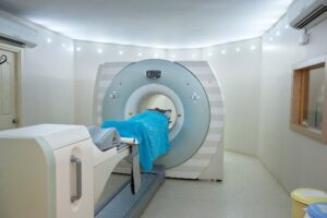
- Imaging studies are helpful in evaluating patients who have symptoms suggestive of peritoneal carcinomatosis. Patients have either a CT scan or MRI of the abdomen and pelvis to confirm the presence of new or recurrent disease in the peritoneum. These imaging studies are done to assess the extent of disease in the abdomen. PET-CT can also be done to determine if disease has spread outside the abdominal cavity, such as the lungs.
- Laparoscopy is performed to confirm the diagnosis and determine if a patient is a candidate for cytoreductive surgery and HIPEC. Laparoscopy is a minimally invasive procedure where, through small incisions, samples of tumor (biopsies) are removed and sent to pathology to confirm a diagnosis. Fluid in the abdominal cavity also can be sampled to look for the presence of cancer cells. This procedure is also used to evaluate the extent of disease in the peritoneal cavity and to calculate the peritoneal carcinomatosis index (PCI) which is used to determine if a patient is a candidate for cytoreductive surgery and hyperthermic intraperitoneal chemoperfusion (HIPEC).